
Repeated Embryo Loss in Mares is the Result of Retarded, Small for Age, Embryonic Vesicles.
By Professor John Newcombe, BVetMed, MRCVS
A Normal Foetus and then a Foal Will Eventually Result When a Normal Embryo is Produced: A Case Report
ABSTRACT

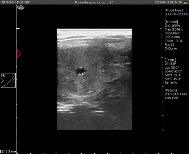
5.5 mm diameter conceptus – normally around 10 days (image not related to the case study)
A twelve-year-old maiden/barren part-TB mare with otherwise limited reproductive history, was sent to an equine fertility clinic for on-site natural mating with an Irish Draught stallion. She was monitored twice daily with ultrasound to determine the time of ovulation and again from Day 12. Embryonic vesicles were measured accurately with ultrasound using electronic callipers. The mare was covered/mated naturally at ten (10) oestrous periods over two breeding seasons by the same stallion. She conceived on seven occasions and returned to oestrus on three. Embryo failure occurred in six of the seven pregnancies. Following the 10th mating, the pregnancy survived, and a foal born. No medical intervention was used to assist embryo survival. Pregnancies were found to have failed by 35, 25, 17, 22, 23 and 21 days sequentially. Embryonic vesicles were considered to be of crucially small diameters for gestational age at each examination on the 6 occasions of failure. They ranged from 2 to 5 mm on Day 14, and from 9 to 14 mm on Days 17/18. Only once did a vesicle grow to more than 18 mm (22 mm) before failure. From an extensive data base held at the Clinic of embryo diameters from the ages of 11 days, the mean diameters of the six failed embryos were smaller than normal by the equivalent in mean growth retardation of 4.0, 3.67, 4.33, 3.67, 3.67, 4.0, 4.33, 5.5, 6.5, 7.0 and 9 days on Days 12 to 22 respectively. On the final and successful cycle, the embryonic vesicle was smaller than the average diameter on Days 12 to 17 and Day 22, representing a growth retardation of the equivalent of <3, 2.5, 2.5, 2.5, 3.0, 3.5 and 4.0 days respectively. The embryo proper was not detected on Day 20 and was small when first seen on Day 23 (c. 2 mm), representing an estimated retardation in development of 3 days. Its development on Day 30, with the allantois occupying approximately 25-30% of the conceptus represented one to two days retardation. The mare subsequently foaled normally.
Keywords: Mare; Repeated Early Embryo Loss; Small for Age Vesicle
Introduction
Embryo loss has been variously shown to be age related (Davies-Morel, et al.1, Rose et al.2) although it is now well accepted that sporadic pregnancy failure may often be the result of Karyotype abnormalities. Embryonic aneuploidy is the leading cause of miscarriage in older women resulting from either faulty meiotic division or gamete chromosomal fusion and has recently shown to be more frequent in older mares (Rizzo, et al.3). Repeated early embryo loss in certain individual mares however has often been ascribed to an adverse uterine environment and various strategies have been forwarded for its prevention. Notably progesterone therapy was and still is, thought to help. Rarely does luteolysis accompany embryo loss, nor is there any general evidence of insufficient luteal support (Irvine, et al. 4). The value of this therapy is enhanced when ad hoc treatment is successful notwithstanding that the next cycle might have been successful with- out treatment! No control study has ever shown that progesterone therapy will reduce the incidence of embryo loss. A recent analysis found no beneficial effect of administering altrenogest in early pregnancy (Cuervo-Arango, et al.5) where embryo transfer pregnancy rates of > 90% and embryo loss rates of <10% were achieved without altrenogest therapy. However very occasionally, an apparently normal vesicle is lost following spontaneous luteolysis at 14/16 days. Some of these can be saved by immediate progesterone therapy at the first signs of luteal regression (significant oedema throughout the uterine lumen and a hypoechoic corpus luteum) (Newcombe: unpublished). The use of 40 mcg buserelin given 10-11 days after ovulation (Newcombe, et al.6) or 10 days after post-ovulation insemination (Unger et al 20027) has shown to significantly improve pregnancy rates and embryo loss rate although the mechanism for this effect remains unclear (Newcombe, et al.6). While instances of embryo failure may be shown to be associated with retained post-ovulatory fluid or even fungal infection (Newcombe8), the majority of losses occur in the absence of other clinical signs. Where the time of conception is known with some accuracy, embryos which appear distinctly small for gestational age are now recognised as often associated with failure (Ball9).
Case History
12-year-old barren/maiden part-TB mare. Previous reproductive history was limited but suggestive of previous embryo failure.
Material and Methods
The mare arrived at the Clinic for routine on-site natural mating. She was examined twice daily to determine a suitable opportunity for covering and to determine the time of ovulation. The mare was mated naturally on each occasion by the same Irish Draught stallion. Examinations were then made from Day 12 onwards for pregnancy. The embryonic vesicle when visualised was measured with electronic callipers from two right-angle diameters. Non-pregnancies returned to oestrus spontaneously and pregnancy failures were returned to oestrus by treatment with 125-250 mcg a PGF2a analogue d-cloprostenol. No pharmacological or other treatments were given during any of the pregnancies.
Results

Table 1
The mare was covered naturally at ten (10) oestrous periods over two breeding seasons by the same stallion. She conceived on seven occasions and returned to oestrus on three. Embryo failure occurred 6 times. On the 10th covering, the pregnancy survived, and a foal born. No medical intervention was used. Pregnancies were found to have failed by 35, 22, 17, 22, 23 and 21 days sequentially. Embryonic vesicles were considered to be crucially small for gestational age on all 6 occasions of failure, with diameters ranging from 2 to 5 mm on Day 14, and from 9 to 14 mm on Days 17/18. Only once did a vesicle grow to more than 18 mm (22 mm). Spontaneous luteal regression with re- turn to oestrus occurred on two occasions resulting in inter-ovulatory intervals of 22 and 23 days. The corpus luteum persisted on the other 4 occasions. Mean diameters of failed embryos on Days 12 to 22 were smaller than normal by 6.44, 9.0, 12.0, 12.8, 13.5, 13.1, 11.6, 10.6, 10.3, 9.8 and 8.0 mm on Days 12 to 22 respectively. This represented the equivalent in mean growth retardation on Days 12 to 22 of > 4, >4, >4, 3.5, 3.5, 4.0, 4.5, 5.5, 6.5, 7.0 and 9 days respectively (Table 1).
On the final and successful cycle, the embryonic vesicle was small- er than the average diameter on Days 12 to 17 and Day 22 by 5.9, 7.8, 8.7, 10.1, 10.5, 10.3 and 3 mm respectively representing a growth retardation of < 3, 2.5, 2.5, 2.5, 3.0, 3.5 and 4.0 days respectively. The embryo proper was not detected on Day 20 and was small when seen on Day 23 (c. 2 mm), representing an estimated retardation in development of 3 days. Its development by Day 30, with the allantoic cavity occupying approximately 25-30% of the conceptus, represented one to two days retardation. Table 1 shows the mean diameters of viable pregnancies in mares examined three times daily for ovulation, and three times daily from 10 days of pregnancy (unpublished data).
Discussion
Embryonic vesicles which are small for gestational age at the first identification, remain retarded including the development of the embryo proper and the first appearance of the allantoic cavity. (Newcombe, et al.10). The degree of retardation appears to remain the same throughout the early development almost as if either the conception date was incorrect, or the conception was to an unobserved later asynchronous ovulation. While there often exists a degree of retardation (or slight advancement) compared with the mean of normal viable pregnancies, there appears to be critical degree of retardation beyond which the pregnancy is non-viable. In this authors experience, vesicles which appear more than 3 days retarded in the first 2 to 2.5 weeks of pregnancy rarely if ever go to term. Beyond 15-16 days, growth is too variably accelerated for comparison with the mean of normal. The greater the degree of retardation, the earlier pregnancy failure will probably occur. In the only three exceptions known to the author to this “more than 3/4 days” rule, all three foals were born alive but died after birth. One succumbed to the Tetralogy of Fallow (Newcombe 2000) but the history of the other two was incomplete. The author has known of several other pregnancies that appeared to be relatively retarded, seemed to be apparently normal at about 40 days, but which were known to have either aborted or failed to foal.
In this Case Report, the time of ovulation and of conception was known, and therefore the age of the embryo was known to within +/- 6 hours (assuming conception always occurs directly after ovulation). However, when examinations to determine the time of ovulation are made only every two days, the embryo age is only known to +/- one day, although a more accurate estimation is possible from the appearance of the pre-ovulatory follicle and/or the appearance of the ovulatory area/CL, estimation of the degree of any growth retardation is therefore less accurate. For example, a vesicle of say 3.7 mm which ap- pears to be 3 days small for its 14 days of age (normal mean 11.6 mm), may actually be only 13 days and only 2 days smaller than the mean, well within the normal range of variability. Not unusually, in a set of synchronous twins, one is smaller for age than the other. When left, even if bilaterally located, the smaller embryo, if significantly smaller, will fail leaving a viable single pregnancy (Ginther11). When ultrasound was in its infancy, practitioners soon learned that reduction of the larger (easier) vesicle often resulted in eventual pregnancy failure, while reduction of the smaller was very much more successful (McKinnon12). An analysis of field data showed that by reduction of the smaller vesicle, significantly more otherwhile twin pregnancies went to term than did single ovulation derived pregnancies (Davies-Morel, et al.13). This was because small for age singletons are not reduced and although they survived beyond the stages of early pregnancy diagnosis, failed subsequently, whereas the larger of twin embryos were mostly normal size for age and more likely to survive.
This case history and other cases seen by the author in the field, illustrate that mares which repeatedly conceive embryos which fail, may yet still conceive a viable pregnancy and foal eventually. A similar case was in a young maiden thoroughbred mare which conceived on 5 occasions at six mattings in a single breeding season. The pregnancies failed on five occasions all before 21 days. At the last mating she conceived and carried a foal to term. All 5 failing embryos were considered small for age (Newcombe unpublished, Newcombe14-16, ).
Conclusion
Embryonic vesicles which are small for age at first identification remain retarded including the development of the embryo proper. There appears to be a critical degree of retardation above which, the pregnancy becomes non-viable. Based upon the diameters recorded during the first 2 to 2.5 weeks of pregnancy, it appears that retardation of more than 3 to 4 days rarely if ever, results in a viable pregnancy. While various unproven therapies are still pursued to treat mares with repeated embryo failure, this case shows that the aetiology of early embryo failure must be embryonic rather than uterine environment. This and other similar cases illustrate that certain mares have a higher risk of small for age embryos probably due to aneuploidy. However, now and then, a ‘good’ oocyte is produced which can align its spindle and separate its chromosomes correctly. So despite multiple pregnancy failures, it is probable that sooner or later, a viable embryo will be produced with the good chance the pregnancy will go to term.
References:
1: Davies-Morel MCG, Newcombe JR, Swindelhurst JC (2005) The effect of age on multiple ovulation rates, multiple pregnancy rates and embryonic vesicle diameter. Theriogenology 63: 2482-2493.
2: Rose BV, Chang YM, Wathes DV, Verheyen KLP, De Mestre AM (2018) Factors that modify the risk of early pregnancy loss and early conceptus growth in thoroughbreds. J Equine vet Sci 66: 179.
3: Rizzo M, Kopps GJPL, Deelen C, Beitsma M, Cristarella S, et al. (2018) Compromised Spindle assembly check-point function in oocytes from aged mares impairs correct chromosome alignment. J Equine vet Sci 66: 177.
4: Irvine CH, Sutton P, Turner JE, Mennick PE (1990) Changes in plasma progesterone concentrations from Days 17 to 42 of gestation in mares maintaining or losing pregnancy. Equine Veterinary Journal 22: 104-106.
5: Cuervo-Arango J, Claes AN, Stoute TAE (2019) In vitro-produced horse embryos exhibit a very narrow window of acceptable recipient mare uterine synchrony compared with in-vivo derived embryos. Reproduction, Fertility and Development 31: 1904-1911.
6: Newcombe JR, Peters AR (2014) The Buserelin Enigma: 2014 How does treatment with this GnRH analogue decrease embryo mortality. Journal of Veterinary Science and Technology 5: 1.
7: Unger C, Schneider F, Hoppen HO, Becker F, Nurnberg G, et al. (2002) Pregnancy rates in mares after application of gonadotrophin releasing hormone analogue, Buserelin, in warmblood mares. Theriogenology 58: 635-638.
8: Newcombe JR (1997) The effect of incidence and depth of intra-uterine fluid in early dioestrus on pregnancy rate in mares. Pferdeheilkunde 13: 545.
9: Ball BA (2011) Embryonic loss. In: Equine Reproduction by McKinnon, Squires, Vaala and Varner. Wiley-Blackwell, Chichester, West Sussex UK. PO19 8SQ 2: 2327-2336.
10: Newcombe JR, Cuervo-Arango J (2012) The relationship between the positive identification of the embryo proper in equine pregnancies aged 18 to 28 days and its future viability. J Equine vet Sci 32: 257-261.
11: Ginther OJ (1989) Twin embryos in mares ll. post-fixation embryo reduction. Equine Veterinary Journal 21: 171-174.
12: McKinnon AO (2007) Twin reduction techniques. In: Current therapy in equine reproduction. Saunders Elsevier, St. Louis USA, pp. 357-337.
13: Davies-Morel MCG, Newcombe JR, Lauber M (2012) Manual reduction of multiple embryos in the mare. The effect on subsequent pregnancy outcome. The Veterinary Journal 192: 322-325.
14: Newcombe JR (2000) Embryonic loss and abnormalities of early pregnancy. Equine Vet Edu 12: 88-101.
15: Newcombe JR (2004) The relationship between the number, diameter and survival of early embryonic vesicles. Pferdeheilkunde 20: 214-220.
16: Unger C, Schneider F, Hoppen HO, Becker F, Nurnberg G, et al. (2002) Pregnancy rates in mares after application of gonadotrophin releasing hormone analogue, Buserelin, in warmblood mares. Theriogenology 58: 635-638.
© 2024 Professor John Newcombe, BVetMed, MRCVS