Evidence that embryo migration (translocation) is not always if ever, necessary for the successful signalling or reception of the Maternal Recognition of Pregnancy (MRP) in the mare
A Case Study
By tracking the movements of the embryonic vesicle throughout the uterine lumen using repeated examinations with ultrasound at one or two hourly intervals from embryo age 10 days until embryo reduction by compression at age 12.3 days, subsequent examinations of the mare were able to determine whether reduction at this time resulted in luteolysis or the Maternal Recognition of Pregnancy (luteostasis)
Materials and Methods
 Anna: Ovulated 4.30h [hours] (+/- 4h) (between 24.30 and 8.30) January 19th 2022
The first examination post-ovulation at 8.30h was deemed to be Interval 1, Embryo age 4h.
First examined for pregnancy from 8.40h January 29th. Interval 31. Embryo age 10 days 4 hours and diameter 3.2 mm. Further examinations were made every hour (Day 10), every two hours (Day 11) and every hour (Day 12)
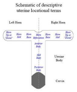
The uterine lumen was arbitrarily divided into 10 locations. 3 locations in the body (anterior, mid and posterior) and 3 in the horn thirds (base, mid and tip or distal) as per Ginther but also included the bifurcation as a distinct location. Movement from one to the adjacent location was 1 unit of migration. Where the embryo position appeared to be between two locations, it was recorded as, for example, mid/anterior body. Movement from a location to a between location was 0.5 units
Results
Day 10. 14 examinations made every hour between 8.40h and 1.00h found the embryo had moved a total of only 3 units (0,2 units/per ex) solely between the mid and anterior body
Day 11. 10 examinations made every 2 hours between 8.30h and 1.30h (Jan 30th) found the embryo had moved a total of 1.5 units (0.15 units/exam) between the anterior body and mid/posterior body
Day 12. At the next examination 8.45h (Jan 31st) , the embryo had moved overnight (8h) from the anterior body to the mid right horn.
A further 8 examinations made between 8.45h and 15.15h (Jan 31st) found the embryo had moved a total 3 units (0.38 units/exam) between the bifurcation and the mid right horn
However, within the interval of 1 hour, the embryo moved from the base of the right to the base of the left horn at which time it was manually reduced at Interval 37.5 (296 hours postovulation)
The mare subsequently entered a period of luteostasis
Embryo reduction had been performed previously in this mare in 15 pregnancies.
When previous reductions were performed at Intervals 32, 33, 34, 35, 36, 36, 37, 37.5, 38, and 38, luteolysis resulted due to of failure to receive the MRP. Reductions at 37.5, 38, 38, 39 and 39 however resulted in luteostasis. It seems that there is critical time for this mare, within a period of 8 hours between Interval 37.5 (296h +/- 4h)) and Interval 38.5 (304h +/- 4h) before which luteolysis results and after which luteostasis results.
Discussion
On this occasion, embryo reduction at Interval 37.5, may or may not have followed by luteostasis
However luteostasis did follow despite only 2, 1, 0, 0, 1 and 2 location changes in the six 8 hour periods up to the 8 hours immediately before reduction. In the last 7 hours before reduction 5 location changes occurred between the bifurcation and the mid right horn (5 location changes on Ginther’s scale)
Resume
The embryo remained in the uterine body for 2 days with also no migration
After 48 hours it had moved into the right horn and oscillated between the mid right and the bif/base right horn for 6.5 hours.
Then within 1 hour it had moved into the base of left horn where it was reduced
The embryo was NEVER seen in either the posterior body, the tip (distal third) of the right horn nor in the mid or tip or the left horn
It was only seen on two occasions in the mid right horn and once in the base of the left horn
Initial (Day10/11) migration was minimal and seemingly had no influence on the signalling of MRP
If we assume the embryo must visit at least a portion of both uterine horns, then it must in this case, have been ‘recognised’ by the endometrium within a short time interval , possibly as short as 1 hour since it was only in the left horn (base) for no more than 1 hour and probably less, before reduction. That is unless, the conceptual remains ie trophoblast and fluid, continue to deliver the signal. Studies by Wilsher suggest that this possibility seems unlikely
Had reduction been performed either 2 or even 1.5 intervals (16 or 12 h) earlier, the MRP would not have been received since all the reductions done at or before Interval 37 resulted in luteolysis. As it was, reduction at Interval 37.5 could have resulted in luteolysis since on two occasions reductions at Interval 38 had resulted in luteolysis
Conclusion
Continued and extensive embryo migration throughout much of the uterine lumen is unnecessary for MRP. Embryo presence in any part of both horns may be needed for a matter of hours. Alternatively migration of the embryo is not necessary for the recognition of the embryo, purely its presence!
© 2022 Professor John Newcombe, BVetMed, MRCVS


