Ultrasound of the Normal and Abnormal Ovulation in the Mare
By Professor John R. Newcombe B.Vet.Med., MRCVS
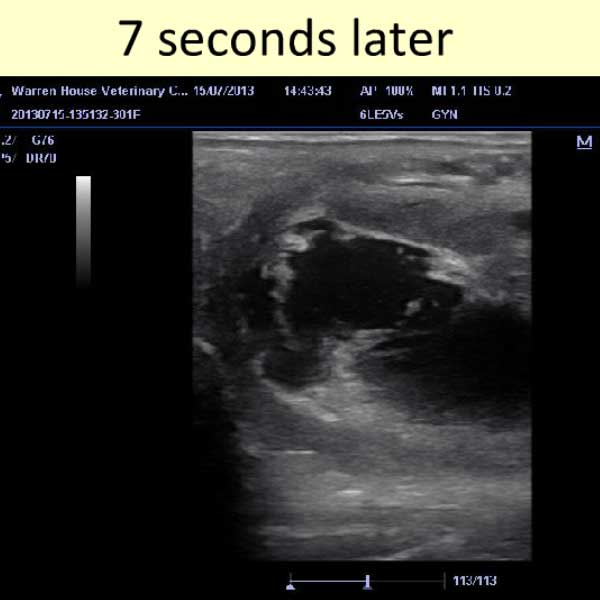
The veterinary practitioner and lay ultrasonographer are often faced with ovarian images which have the potential to perplex, as they fall outside what the imagination has created to be a “normal” appearance for the stage being evaluated. In this presentation of ultrasound images John Newcombe takes us through the pre-ovulatory stages of a follicle, ovulation itself and the development of the corpus haemorragicum (“CH”) and/or corpus luteum (“CL). In addition, abnormal ovulations are imaged and reviewed, including Haemorrhagic Anovulatory Follicles (“HAF” – also known as “Anovulatory Haemorrhagic Follicles” or “AHF”) and Luteinized Unruptured Follicles (“LUF”) as well as an evaluation of a large variety of different appearances of the normal, along with explanations.
This PowerPointTM presentation was first shown at the 2013 BEVA Convention. It has been converted to a video for use on this site. We have attempted to provide adequate time to view each slide, but if more time is required, you will need to pause the video.
© 2014, JR Newcombe and Equine-Reproduction.com, LLC
Use of article permitted only upon receipt of required permission and with necessary accreditation.
Please contact us for further details of article use requirements. Other conditions may apply.


