
Vitrification of Larger Embryos
Successful vitrification of larger embryos – those >300µm – has typically not been achieved previously without use of the comparatively new procedure of puncture. Prior to development of that technique, best success was seen with smaller embryos, which has necessitated flushing on day 6 or 6½ post-ovulation. This in turn required closer monitoring to determine exact time of ovulation, and also could result in reduced recovery rate either because the embryo has not descended into the uterus at the time of the flush, or it was either not recovered, or found during the search. The puncturing procedure when performed, was commonly done with the use of micromanipulators and needles such as are used during ICSI. There has been success reported with the use of a manual puncture process, but the searched for “golden egg” has been the successful vitrification of larger embryos without puncture.
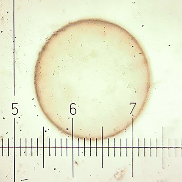
This one is clearly too large!
Kovacsy et. al. reported a new method for vitrification of day 7-8 post-ovulation embryos ranging in size from ≤300 µm (8) to >500µm (6), with an additional 16 in an intermediate-sized group. A human embryo vitrification kit was used (Kitazato) with the embryos initially placed in an equilibration medium containing 7.5% Ethylene Glycol and 7.5% DMSO at room temperature for 15 minutes. They were then transferred to a vitrification solution for up to 90 seconds, during which they were loaded onto a Cryolock device, capped, and then plunged into liquid nitrogen.
Subsequently, the embryos were thawed, then placed in two differing osmotic pressure holding media for 1 minute and 4 minutes prior to being placed in a standard holding media for an additional 4 minutes. Within 15 minutes, the thawed embryos were then transferred into recipient mares which were 6 days post-ovulation. The mares were checked three times to confirm a developing pregnancy prior to being given prostaglandin. Two of the mares were kept pregnant until embryonic heartbeat was detected to confirm normal development.
87.5% (7/8) of the <300 µm embryos underwent successful thawing and transfer, and developed an ongoing pregnancy; 75% (12/16) of the intermediate group (>300 to ≤500µm diameter) successfully transferred and developed, although one of 350µm diameter did undergo EED. The four in this intermediate group that failed to survive transfer and develop were of mixed diameters (330, 380, 425 and 500µm). None of the embryos >500µm survived.
Previously, vitrification of larger embryos has been thought to require puncture – or in some cases puncture coupled with aspiration of the blastocoele. In this research, with a surviving pregnancy rate of 79% (19/24) for embryos <500µm in diameter, this is clearly not an absolute requirement. This new technique opens the doors for a simpler – and successful – method of vitrification for larger embryos, including those flushed on day 7 and even possibly some of those being smaller on day 8, without the use of expensive equipment or more complicated techniques.
(Kovacsy S, Ismer A, Funes J, Hoogewijs M, Wilsher S. 2023. Successful vitrification of embryos ≤500µm without puncture or aspiration of the blastocoele. JEVS 125:104656)