
A New Method to Estimate Fetal Age in the Mare
Sometimes there arises a need to estimate fetal age in the mare, for which a breeding date perhaps is unknown. Several methods have been previously identified, such as measurement of the fetal eye[1][2], or biparietal diameter, eye approximated volume, fetal aortic diameter and femur length[3], among others. These methods are of value, however positioning of the fetus in utero to allow accessibility, and in some cases mixed reliability based upon the age at time of evaluation would encourage one to seek additional methods for at the very least, cross reference purposes, particularly through the later stages of pregnancy.
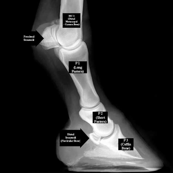
 Renaudin et al. reviewed 10 mares (9 QH and one TB recipient carrying a QH fetus) from 9 months of gestation through to parturition. Ultrasonographic evaluation of the fetal second phalanx (P2 – short pastern), distal metacarpal 3 (MC3 – cannon bone), and the proximal and distal (navicular bone) sesamoids was performed to determine if through measurement or other changes one can estimate fetal age in the mare. Evaluation and measurements were performed at two-week intervals.
Measuring the ossified portion of the length of P2 (the short pastern), after 236 days of gestation, a strong correlation to gestational age was found, to the point where a predictable formula was developed: y=0.1413x minus 24.89 (where y=predicted value of P2 length in mm and x=fetal age).
The appearance of the epiphyses at distal M3 and proximal P2 provided another useful – although slightly less precise – timeline, first being visible at between 260 and 294 days of gestation (mean: 268 days) and 285 and 306 days of gestation (mean: 294 days) respectively. In no instance was closure of the epiphyses seen prior to parturition.
Similarly, the appearance of the sesamoids provided a somewhat predictable but variable timeline for estimate of fetal age, the proximal sesamoids being first observed between 286 and 306 days of gestation (mean: 295 days), and the distal sesamoid (navicular bone) between 300 and 329 days of gestation (mean: 317 days). It must be noted however, that there was a distinct outlier in the timing of the appearance of the sesamoids, with them first becoming visible at 331 days (proximal) and 335 days (distal) of pregnancy in the smallest foal in the group.
This research demonstrated a useful and reliable method to estimate fetal age in the mare in the later gestational period. A particular convenience of the method is that in the normal presentation, the distal forelimbs will be in a position adjacent to the cervix within this time frame, making it easy to achieve measurements with accuracy.
(Renaudin CD, Wensley FM, Morgan JM, Cassano JM, Spriet M. 2023. Fetal ultrasonography of the distal limb: a new tool to assess equine fetal age and bone development. JEVS 125:104781)
References:
1: Kahn VW, Leidl W. 1987. Die ultraschall-biometrie von pferdefeten in utero und die sonographische darstellung ihrer organe. Dtsch tierarztl Wschr, 94:497–540.
2: Turner RM, McDonnell SM, Feit EM,Grogan EH, Foglia R. 2006. Real-time ultrasound measure of the fetal eye (vitreous body) for prediction of parturition date in small ponies. Theriogenology 66:331–337.
3: Renaudin CD, Kass PH, Bruyas J. 2022. Prediction of gestational age based on foetal ultrasonographic biometric measurements in light breed horses. Reprod Dom Anim. 57:743-753.