

Endometrial Biopsy in the Mare – is one sample enough?
An endometrial biopsy in the mare is a useful diagnostic to determine cellular health of the uterine lining which will be directly responsible for pregnancy maintenance once placentation has occurred. The biopsy score is correlated to the likelihood of live foal production by use of different scales, such as that of Kenney and Doig (modified) which offers 4 reporting categories (3 numerical, one sub-divided), correlated to statistical live foal production ranging from <10% to 80-90%.
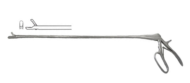
Jackson Biopsy Forceps
The equine uterus is a comparatively large organ, so it has been questioned whether a single-sample endometrial biopsy in the mare would be representative of the condition of the entire uterus. This has been reviewed previously, with mixed results. It has been reported that distribution of endometrial glands is uniform throughout the uterus [1] which is suggestive that there may be a uniformity of results. At least one University Teaching Hospital has presented work suggesting 92% accuracy with a single sample, increasing only to a 96% accuracy when 10 samples are taken [2]. Other published work has also supported this suggestion [3][4]. This is in contrast to other reviews which suggested a lack of uniformity of findings throughout the uterus [5]. The question therefore persists – does one sample site provide adequate answers for an endometrial biopsy in the mare?
Muderspach et al. re-evaluated this question with a particular emphasis on differences in specific degenerative changes between sampling sites. As an adjunct, animal age, breed and estrous cycle stage differences were considered.
Postmortem samples were taken from two sites – the left and right horn – from 82 mares. After staining, these were evaluated, and a score applied to different conditions (periglandular fibroblasts, glandular nests per view, dilated glands per view, degree of glandular dilation, excessive lymphatic vessels, and lymphatic lacunae). In addition, the standard modified Kenney and Doig classification was applied to each sample.
Under half of the paired specimens received the same “Kenney and Doig” scores on both samples (49%). In 46%, the variations were a single category variation (in either direction), while in the remaining 5%, the variation was 2 categories. Disagreement in the scores for the individual conditions varied from 10% (glandular nests) to 38% (excessive lymphatic vessels). In mares under the age of 20 however, the cumulative scores for these changes did not differ between horns in individual mares.
Based upon the findings of these samples and scores, the recommendation is given that caution should be used when evaluating a single-sample endometrial biopsy in the mare, in particular in mares over 20 years of age. To improve accuracy of the endometrial biopsy in the mare, two biopsy samples are recommended.
(Muderspach ND, Agerholm JS, Troedsson MHT, Ferreira-Dias G, Christoffersen M. 2023. Are degenerative changes in a single endometrial biopsy representative for the endometrial state? JEVS 125:104734)
References:
1: Blanchard TL, Garcia MC, Kintner LD, Kenney RM. 1987. Investigation of the representativeness of a single endometrial sample and the use of trichrome staining to aid in the detection of endometrial fibrosis in the mare. Theriogenology 28:445–450
2: Ohio State University, unpublished data.
3: Kenney RM. 1978. Cyclic and pathologic changes of the mare endometrium as detected by biopsy, with a note on early embryonic death. J Am Vet Med Assoc 172:241-262.
4: Kenney RM, Doig PA. 1986. Equine endometrial biopsy. In: Morrow D (ed) Current therapy in theriogenology. 2 ed., Saunders, Philadelphia, pp 723-729.
5: Keller A, Neves AP, Aupperle H, Steiger K, Garbade P, Schoon HA, Klug E, Mattos RC. 2006. Repetitive experimental bacterial infections do not affect the degree of uterine degeneration in the mare. Anim Reprod Sci 94:276-279.