Diagnosis of Placentitis – Are Inflammatory Markers Useful?
Placentitis is – literally and practically – “inflammation of the placenta” (“itis” as a suffix indicates inflammation). Use of assays for inflammatory markers in the mare – in particular Serum Amyloid A (SAA) – have been previously suggested as possible diagnostic tools for use in the identification of placentitis. Inflammatory markers including SAA been recognized however as possibly being non-specific to placental inflammation should other inflammatory conditions exist in the animal. Previous research evaluated SAA levels in mares with induced placentitis, and confirmed elevated SAA levels to be present [1]. Feijo et al. revisited this finding, reviewing immunological, inflammatory, and hormonal parameters, including SAA, alpha-fetoprotein (AFP), progesterone and estrogen in mares identified as having an early onset of naturally occurring placentitis.
Two large herds of mares were monitored by reproductive (ultrasound) examinations between 180 and 320 days of gestation also underwent blood sampling at the same time. 12 mares were identified early diagnosis of placentitis as evidenced during ultrasound examinations, presenting with one or more symptoms such as cervical relaxation, placental thickening, abnormal fluid presence, placental edema, and/or placental separation. 12 comparable (age-matched) non-placentitis mares were used as controls.
On the day of being identified by ultrasound with the early onset placentitis, blood draws were performed in affected and the non-placentitis control mares. The results of assays from these samples were then compared to the results from the previous routine monthly assay from the same mares, when no ultrasonographic abnormalities had been identified, as well as the control mares. The duration between these two draws was approximately 40 days (“unaffected” draw: 227±6 days; early-onset or control draw: 267±5 days).
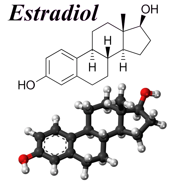
No differences were found in the inflammatory markers SAA or AFP between the two timeframe samples in either group (affected or control). Similarly, no difference was identified in plasma progesterone levels, nor the progesterone/estradiol 17ß ratio. A decline in plasma estradiol 17ß levels was however noted, dropping from levels of 498±35 pg/mL to 408±35 pg/mL, which was considered to be a useful marker for early diagnosis of placentitis, and worthy of further evaluation.
Our comment: As has been previously noted, SAA in particular, although a convenient assay, was not a specific diagnostic marker for placentitis, and for the animals in this research its reliability for early diagnosis of placentitis was non-existent. Extreme caution should therefore be used in cases where SAA is being used as a potential diagnostic for placentitis. The decline in the level of estradiol 17ß is a potentially useful confirmation of clinical evidence suggested by others as being an indicator of possible placentitis and/or fetal compromise, as the fetus is the producer of elevated levels of estrogens (and other hormones) during gestation.
(Feijo L, Wolfsdorf K, Canisso IF, Felippe J. 2013. Diagnosis of imminent placentitis in mares. JEVS 125:104766)
References:
1: Coutinho da Silva MA, Canisso IF, MacPherson ML, Johnson AEM, Divers TJ. 2013. Serum amyloid A concentration in healthy periparturient mares and mares with ascending placentitis. Equine Vet J 45(5):619-24.


