
Indications of Impending Ovulation in the Mare
By Jos Mottershead
Predicting ovulation in the mare can be difficult, as they have the capacity to be notoriously unpredictable in their timing! This can lead to frustration, aggravation and distress in the owners and attending veterinarians! What signs may one hope to see that could provide some mile markers along the way?
Let’s lay out some ground rules, but remember that mares make a point of not following rules, so there will always be exceptions! Note that we are referring to full-sized horses and ponies in this outline, not miniature horses.
- Follicular diameter must be determined by averaging the widest and narrowest parts visible per ultrasound. Variations between the two are likely, meaning that palpation alone will tend to result in less accurate diameter estimations;
- Most mares are less likely to ovulate with a follicle that is smaller than 35 mm;
- Double ovulations with follicles on the same ovary are likely to occur with smaller diameter follicles – possibly less than 35 mm;
- Follicular growth is approximately 3-5 (three to five) mm per day;
- Uterine edema – an important indicator of estrus – peaks around 36 hours prior to ovulation in healthy mares. Mares that have edema that persists closer to or after ovulation are more likely to have delayed uterine clearance issues;
- A change in regularity of follicular shape visible per ultrasound – with a tendency towards a “teardrop” or “guitar pick” shape – is likely to be seen within the 24 hours prior to ovulation in about 85% of preovulatory follicles;
- A thickening of the follicular wall visible per ultrasound – evidenced by a thicker white line surrounding the follicle (especially visible on the lower margin) – is likely to be seen within 24 hours prior to ovulation in about 85% of preovulatory follicles;
- Ovarian sensitivity to touch upon palpation per rectum may be demonstrated within the 12 hours prior to ovulation in some mares (make sure you differentiate this from the mare that is always sensitive upon palpation. The “ovarian sensitivity” marker refers only to mares that have a change in response);
- Most follicles will become softer upon palpation per rectum within 6-12 hours prior to ovulation. While more easily identified by palpation, application of slight pressure with the rectal probe of an ultrasound that results in a flattening of the upper surface of the follicular image may also be indicative of follicular softening;
- Most follicles will reduce slightly in average diameter at some point during the 6 hours prior to ovulation;
- There may be slight evidence of white specks in the follicular fluid within the 12 hours prior to ovulation, which should not be confused with indications of an anovulatory hemorrhagic follicle;
- A visible “leaking” of the follicular fluid outside the main portion of the follicle per ultrasound is indicative of immediately impending or ongoing ovulation;
- Breeding up to 6 hours post-ovulation will typically result in similar pregnancy rates as would be seen with pre-ovulatory breeding. It has been shown that pregnancy rates will not be decreased, nor early embryonic death rates increased with breedings up to 16 hours post-ovulation if additional management practices (lavage, oxytocin, antibiotic infusion) are performed (Newcombe JR, Cuervo-Arango J. The effect of time of insemination with fresh cooled transported semen and natural mating relative to ovulation on pregnancy and embryo loss rates in the mare. Reprod Domest Anim. 46(4):678-81).
- Mares may continue to “tease” to a stallion for 12-48 hours post-ovulation. This is not indicative of a problem, and is normal behaviour.
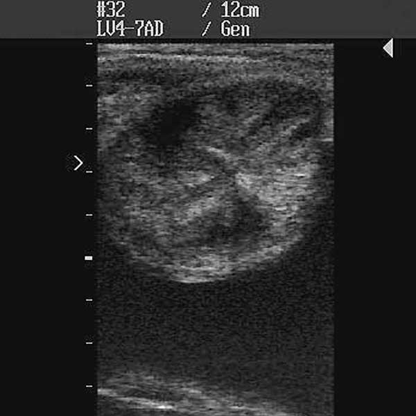
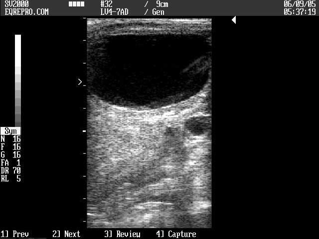
Ultrasonic Images

Cross-section of uterine horn showing pronounced edema per ultrasound

Follicular width and height are not necessarily the same and must be averaged

Double follicles may ovulate at a smaller size, and may feel like a single follicle on palpation
A flattening effect to the top of the follicle may be seen when gentle pressure is applied with the ultrasound probe once the follicle starts to soften prior to ovulation

A distinctive change to a “teardrop” shape may be seen within 24 hours prior to ovulation

Leaking of follicular fluid exterior to the follicle but within the ovary may be seen immediately prior to and during ovulation
All images are from ultrasounds performed with a Digital Sonovet 2000 unit, available from Universal Imaging Inc.
| Follicle: | The structure that forms on the ovary containing the oocyte. It is the follicle that is said to “ovulate”. |
| Ovulate/ovulation: | The process whereby the oocyte is released from the follicle. The follicle ruptures typically at a point close to the ovulation fossa, and the rush of the follicular fluid forces the oocyte towards the infundibulum of the oviduct. |
| mm/cm (millimetres/centimetres) | The commonly used measurements for follicular diameter. 10 mm = 1 cm, hence 35 mm = 3.5 cm |
© 2006 Equine-Reproduction.com, LLC
Use of article permitted only upon receipt of required permission and with necessary accreditation.
Please contact us for further details of article use requirements.
Other conditions may apply.